Open Journal of Pediatrics and Child Health
Something lurks in my baby’s gums
Rita Justo Pereira1*, Pedro Cruz2, Paulo Sousa3 and Luís Gonçalves2
2Pediatrics and Neonatology Department, Private Hospital of the Algarve, Faro, Portugal
3Surgery Department, Private Hospital of the Algarve, Faro, Portugal
Cite this as
Pereira RJ, Cruz P, Sousa P, Gonçalves L (2023) Something lurks in my baby’s gums. Open J Pediatr Child Health 8(1): 001-002. DOI: 10.17352/ojpch.000044Copyright
© 2023 Pereira RJ, et al. This is an open-access article distributed under the terms of the Creative Commons Attribution License, which permits unrestricted use, distribution, and reproduction in any medium, provided the original author and source are credited.Abbreviations
CGCE: Congenital Granular Cell Epulis; MRI: Magnetic Resonance Imaging
Case report
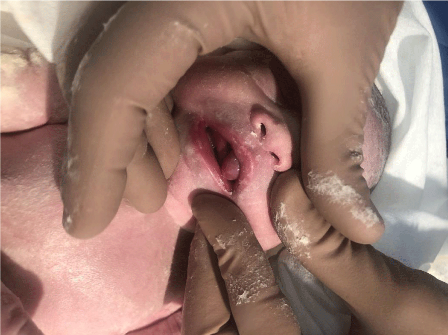
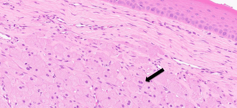
A healthy Caucasian female neonate was born at 38 weeks and 3 days of gestation by elective cesarean delivery. She is the second child of a healthy mother, had a proper antenatal follow-up, and had no fetal anomalies in prenatal ultrasounds. The birth was uneventful with an apgar score of 9/10/10 at one, five and ten minutes respectively, and a birth weight of 2685g (P3-15). The first physical examination revealed a female neonate, covered by vernix caseosa, with a red, regular, 1 cm long, appendicular lesion on the anterior part of the maxillary alveolar ridge (Figure 1). There were no apparent malformations or other abnormal findings on the newborn´s examination. A Congenital Granular Cell Epulis (CGCE) was clinically suspected by its appearance and location, and parents were informed and reassured of the benign nature of the lesion. In the first hours of life, she presented a normal sucking reflex but an incorrect breast latch. There was no evidence of airway obstruction. Parental support was essential to the successful breastfeeding of this neonate. A prompted surgical excision of the lesion was performed on the first day of life, with parental consent. The anatomopathological examination revealed a mass of 12 mm x 10 mm with the subepithelial proliferation of granular cells (Figure 2), consistent with CGCE. A complete and jusxtalesional excision was done using cautery (Figure 3), without any complications in the postoperative period. Since then, the newborn has acquired a good breast latch and maintained breastfeeding on demand. There was no recurrence after one year of follow-up.
Discussion
CGCE is a rare benign tumor of the alveolar ridge most frequently found in the maxilla [1,2], at the canine-incisor region. The mandibular region can also be involved. Etiology remains unclear and there is a female predominance (8:1) [1]. The incidence of CGCE has been estimated as 1/1.000.000, with a few cases reported in the literature [3]. Diagnosis is generally clinically based upon the presence of certain distinctive features such as occurrence only in the newborn, the typical location, and the plexiform arrangement of the capillaries. Although benign, the tumor may vary in size from a few millimeters to several centimeters and can be large enough to compromise a newborn’s airway and feeding [1-6]. It is usually diagnosed at birth (or just after birth), however, large tumors can be detected on antenatal ultrasounds [5]. Oral cavity imaging studies such as MRI are recommended for differential diagnosis.
Treatment consists of surgical excision due to the possibility of interference with breastfeeding and airway obstruction. The excision can be performed under local or general anesthesia, depending on the size and location of the lesion. There may be spontaneous regression in small lesions [1,6].
An anatomy pathology study with histopathological examination of the excised specimen is mandatory for confirming the diagnosis. In this case, the histologic examination confirmed CGCE with no malignant features. A follow-up period is highly recommended because recurrence after surgical removal is possible. It is crucial for Pediatricians to recognize this type of lesion, in order to advise parents of optimal treatment.
- Almiro MM, Santos L, Henriques R, Afonso E. Epúlide Congenita. Acta Pediatr Por. 2015; 46: 273-6. https://doi.org/10.25754/pjp.2015.6623
- Chinnadurai S, Goudy SL. Neonatal airway obstruction: An overview of diagnosis and treatment. Neo Reviews. 2013; 14: e128-37. 10.1542/neo.14-3-e128
- Bosanquet D, Roblin G. Congenital epulis: a case report and estimation of incidence. Int J Otolaryngol. 2009;2009:508780. doi: 10.1155/2009/508780. Epub 2009 Nov 19. PMID: 20130770; PMCID: PMC2809329.
- Siqueira AS, Carvalho M, Monteiro A, Pinheiro M, Pinheiro L. Congenital epulis with auto-resolution: case report. Rev Gaúch Odontol. 2014; 62(3):315-8. https://doi.org/10.1590/1981-86372014000300000131891
- Rodrigues KS, Barros CC, Rocha OK, Silva LA, Paies MB. Congenital granular cell epulis: case report and differential diagnosis. J Bras Patol Med Lab. 2019; 55(3):281-8. https://doi.org/10.5935/1676-2444.20190026
- Kadlub N, Galliani E, Oker N, Vasquez MP, Picard A. Congenital epulis: Refrain from surgery. A case report of spontaneous regression. Arch Pediatr 2011; 18:657-9. doi: 10.1016/j.arcped.2011.03.011
Article Alerts
Subscribe to our articles alerts and stay tuned.
 This work is licensed under a Creative Commons Attribution 4.0 International License.


 Save to Mendeley
